


 16 / 78

110
Ces Urol 2015; 19(2): 106–117
PŘEHLEDOVÝ ČLÁNEK
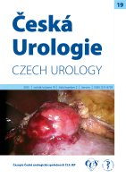
Obr. 3.
Žena ve věku 18 let, s geneticky prokázanou TS (mutace genu TSC1) vyšetřována pro hematurii. Doplněno
CT a následně MRI s nálezem objemného 8 cm velkého AML v dolní třetině pravé ledviny
Fig. 3.
An 18-year-old female with a genetically confirmed TS (TSC1 gene mutation) investigated for haema-
turia. CT was performed and complemented by MRI with a finding of an 8-cm large AML in the lower third of
the right kidney
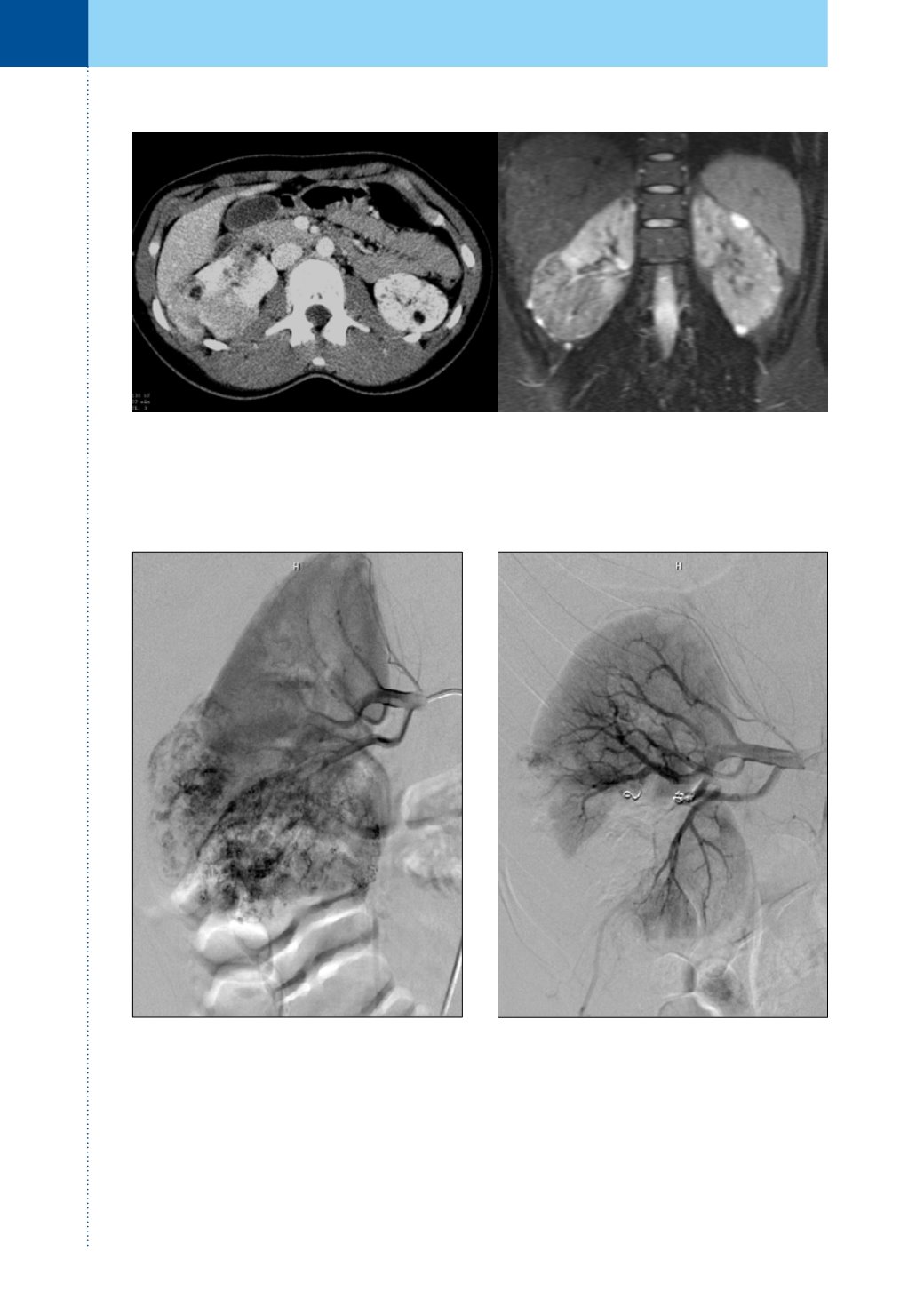
Obr. 3a.
Byla provedena renovasografie s patologic-
kou vaskularizací dolního pólu pravé ledviny a četnými
aneuryzmaty velikosti do 5 mm
Fig. 3a.
Renovasography was performed, showing
a pathological vascularization of the lower pole of
the right kidney and numerous aneurysms with sizes
of up to 5 mm
Obr. 3b.
Vzhledem k tomu, že resekční výkon by
byl obtížně technicky proveditelný, byla provedena
arteriální embolizace PVA částicemi 200 µ, absolutním
alkoholem a spirálami
Fig. 3b.
Given the fact that a resection procedure
would be technically challenging, arterial emboliza-
tion with 200µm PVA particles, absolute alcohol, and
coils was performed