
Ces Urol X:X
Verification of renal ischemia during selective clamping of the renal artery branch during robot assisted resection of a renal tumor using ICG and NIR imaging – video
- 1 Urologická klinika LF UK a FN Plzeň
- 2 Klinika zobrazovacích metod LF UK a FN Plzeň
- 3 Šiklův ústav patologie, LF UK a FN Plzeň
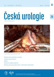
Objective: Verification of renal ischemia during renal artery clamping in minimally invasive renal tumor resection is possible based on the appearance of the kidney, using Doppler ultrasonography and indocyanine green (ICG) administration with narrow light spectrum application (NIR (near infrared) imaging/fluorescence). In most cases, the first two methods are sufficient; we use ICG very sparingly, only when selecting clamping of multiple arteries or branches of the renal artery. The video shows the use of ICG in robotic resection (RR) of a kidney tumor with a duplex renal artery. Abstract: ICG with NIR imaging technology has been available since July 2018. Between July 2018 and May 2020, it was used in 19 (15.2%) of 125 laparoscopic resections – Olympus Visera Elite CLV-S200-IR equipment. For RR, it was used in 9 (2.4%) out of 340 cases between June 2020 and October 2025. For RR, we use the FireFly™ technology of the da Vinci Xi system. Technique: After clamping the renal artery (usually selective clamping of only some of the branches), 1.25 mg of ICG (Verdye®) is administered intravenously and the display is switched from classic mode to NIR fluorescence imaging (FireFly™). The video shows the described methodology for selective clamping of the duplex renal artery during RR.
Results: In all 28 cases, the methodology clearly showed the ischemic part of the kidney.
Conclusion: We usually use Doppler imaging to verify the effectiveness of ischemia during clamping of the renal artery or its branches. In highly selected cases (especially multiple arteries or clamping of only one branch of the artery), ICG administration with NIR imaging can be used.
Keywords: kidney tumor – resection – laparoscopy – robot – ICG
Received: November 2, 2025; Revised: November 2, 2025; Accepted: November 18, 2025; Prepublished online: January 21, 2026
File not found
Verifikace ischemie ledviny při selektivním klampování větve renální tepny při robotické resekci tumoru ledviny pomocí ICG a NIR zobrazení – video
